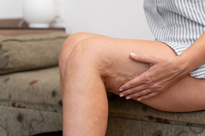
Varicose veins, varicose veins, varicose veins(from the Latin varix, varicis - dilation, swelling in the veins)- persistent and irreversible change in the veins, characterized by:
- uneven increase in the lumen and length of the veins,
- the formation of nodules in areas of thinning of the veins due to pathological changes in the venous walls, their thinning, stretching, decreased tone and elasticity,
- functional insufficiency of venous valves and impaired blood flow.
Varicose veins are a fairly common disease. Varicose veins and their complications are diagnosed in 25% of the population, with women suffering 2 to 3 times more often than men. In women, the first signs of the disease are often observed at a young age, most often associated with pregnancy and childbirth. At more advanced ages, there is an increased incidence in both men and women, and the frequency of complicated forms increases. At the age of 70, the disease occurs 6-10 times more often than at the age of 30. However, recently, the manifestation of varicose veins has been frequently observed in very young people, even teenagers. Therefore, identifying all possible causes of the disease and preventing venous disorders becomes an increasingly urgent task.
How varicose veins arise and develop
To understand how varicose veins occur, let's briefly explain the physiology of the venous system of the lower extremities. Venous flow in the legs is provided by two interconnected mechanisms - central and peripheral. The central mechanism is associated with the heart, lungs, diaphragm, and the peripheral mechanism is directly related to the functioning of the venous system of the lower extremities that surrounds the vessels of muscles and tissues.
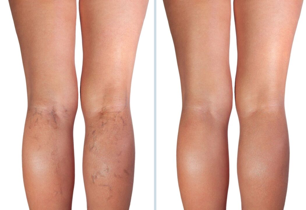
The initial signs of varicose veins are impaired capillary circulation, swelling, a feeling of heaviness in the legs, the appearance of spider veins and spider veins. These changes are reversible, but if preventive measures are not taken in time, problems will increase.
As a rule, more than a year passes from the initial signs of varicose veins of the lower extremities to the formation of varicose nodules and the appearance of pronounced symptoms of varicose veins. Developing gradually, varicose veins lead to impaired blood flow and chronic venous insufficiency. Stagnation of blood in the veins can cause phlebitis (inflammation of the veins), thrombophlebitis (inflammation of the veins with the formation of blood clots), phlebothrombosis (thrombosis with greater inflammation of the veins), non-healing dermatitis (inflammation of the veins). skin), trophic ulcers.
Types and shapes of varicose veins

There are primary (true) and secondary (symptomatic) varicose veins.
Primary varicose veins are an independent disease of the venous system (varicose veins). It develops gradually over several years. Most often, varicose dilatation of the great saphenous vein (70-85%) is observed, less often - of the small saphenous vein (5-12%). In varicose veins, 50-70% of vein damage is bilateral.
Secondary varicose veins are a symptom and consequence of diseases in which there are obstructions to the flow of blood through the deep veins of the lower extremities (post-thrombotic disease, tumors, scars, inflammatory processes, aplasia and dysplasia of deep veins, arteriovenous fistulas, etc. ). Secondary varicose veins are quite rare.
Most often, varicose veins affect the saphenous veins of the lower extremities, which are part of the great saphenous vein system. The branches of the small saphenous vein suffer from varicose veins much less frequently.
Classification of types of varicose veins
Until recently, in our country, doctors classified varicose veins according to different types of classifications. V. S. Savelyev's staged clinical classification was used, reflecting the degree of disturbance of venous circulation in the limb and the body's ability to resist these disturbances and compensate for them, as well as the classification according to the forms of varicose veins and complications due to they caused.
But the main one currently is the international CEAP classification, based on the clinical (C - clinical), etiological (E - etiology), anatomical (A - anatomy) and pathogenetic (P - pathogenesis) characteristics of the disease.
6 clinical classes ("C") are organized according to the increasing severity of the disease, from telangiectasias (TAE) to trophic ulcers.
The etiological section ("E") indicates whether the process is primary or not.
The anatomical part of the classification ("A") divides the venous system of the lower extremities into 18 relatively separate segments, which makes it possible to indicate the location of the affected area of the venous system.
The pathophysiological cut ("P") characterizes the presence of reflux and/or obstruction in the affected venous segment.
Symptoms of varicose veins

Symptoms of varicose veins depend on the stage of the disease, that is, on the degree of changes in blood vessels and disturbances in the venous system. Depending on the stage, a prognosis for the development of the disease can be given.
The initial stage of varicose veins - varicose veins of the 1st degree
At the initial stage, when the pathology of the veins is not yet clearly expressed, visible signs of varicose veins may be absent. Patients complain of a feeling of heaviness and discomfort in the legs, very rapid fatigue, a feeling of heat, paresthesia (numbness, burning, tingling). Symptoms worsen at the end of the day, as well as under the influence of heat - in summer, or when wearing warm shoes indoors in winter. Swelling appears in the foot and ankle, which disappears after a short rest. Occasionally, nocturnal cramps in the calf muscles are possible, but patients attribute them to overwork.
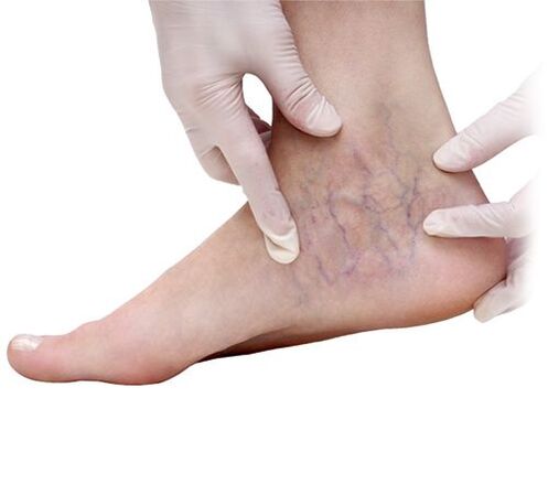
After prolonged physical activity, the veins swell and their network can be easily seen through the skin. They are especially visible in the thighs, legs and feet. The number of these veins and the degree of their expansion may vary. They may be unique and barely noticeable formations on the lower part of the leg, appearing more clearly at night or after physical activity. Also at this stage of varicose veins, spider veins appear.
If at this stage you start the simplest conservative treatment, as well as follow preventive measures, the development of the disease can be prevented by eliminating almost all symptoms.
Symptoms of 2nd degree varicose veins, compensation phase
At this stage of the disease, changes in the large subcutaneous vessels become noticeable. The veins become deformed, swell, the blood flow stops, and noticeable swelling appears in the feet and ankles. Swelling increases with prolonged physical activity in the legs, but disappears after a night's rest. At night, cramps in the calf muscles are common. Paresthesia is observed - temporary loss of sensitivity in the legs, numbness in the legs, burning, "goosebumps". As the disease progresses, pain appears, which intensifies at night.
This phase of subcompensation, as a rule, lasts several years, and at this time the development of the disease can also be stopped if treatment is started in a timely manner. Otherwise, the disease will inevitably progress to a more serious stage.
Symptoms of 3rd degree varicose veins - decompensation stage
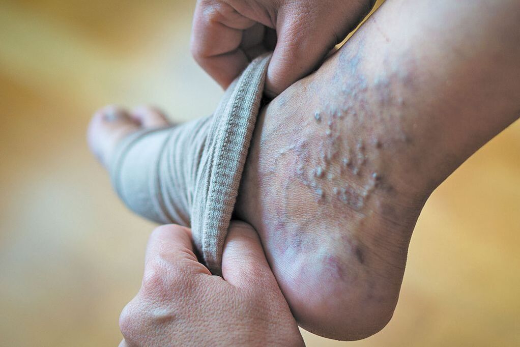
At this stage of varicose veins there is a significant increase in symptoms, the pain and heaviness in the legs are more intense and there is a disturbance in peripheral blood and lymphatic circulation (chronic venous insufficiency). The swelling does not go away even after a long rest and spreads to the lower leg. Patients are bothered by itchy skin. The skin on the legs becomes dry, loses elasticity, the skin is easily injured, loses its ability to quickly regenerate, so wounds take a long time to heal. Brown spots appear on the skin, most often on the inner surface of the lower third of the leg (hyperpigmentation due to subcutaneous hemorrhages).
All these complaints are constant. In the future, complaints of pain in the heart region, shortness of breath, headaches and deterioration in the musculoskeletal function of the affected limb may arise.
Although the decompensation phase is already a very significant manifestation of the disease, with adequate treatment the patient's condition can be maintained at a satisfactory level for a long time, maintaining work capacity and avoiding the transition to the complication phase.
4th degree varicose veins - complications stage
This stage of the disease is characterized by pronounced disorders of venous circulation. Swelling of the legs becomes almost constant, itching of the skin intensifies, and trophic disorders appear on the skin of the leg. Advanced varicose veins are often accompanied by eczema, dermatitis and long-lasting lesions, and as the regenerative capabilities of skin with varicose veins are visibly reduced, even a small wound can develop into a persistent trophic ulcer. The thinned skin and venous walls are easily injured, resulting in extensive bleeding. Damaged soft tissue and open ulcers become entry points for infections.
The most common complications of varicose veins:
- phlebitis - inflammation of a vein;
- thrombosis - formation of a blood clot (thrombus) in a vein, which can lead to blockage of the vessel;
- trophic ulcers - are formed in the place where the affected vein cannot provide sufficient flow of blood from the skin, resulting in disruption of nutrition (trophism) of the tissues.
Varicose veins can be complicated by acute (sometimes purulent) thrombophlebitis.,dermatitis and eczema, bleeding, erysipelas, lymphangitis.One of the most dangerous complications of varicose veins is pulmonary embolism, which can lead to sudden death.
At this stage, it is no longer possible to restore the state of the venous system, we can only talk about preventing new complications and, as far as possible, improving the patient's quality of life.
Causes of varicose veins
There is no single cause for primary varicose veins of the lower extremities. The development of this disease is usually caused by several factors. But all the painful symptoms of varicose veins are associated with structural changes in the tissue of the venous walls of blood vessels and disruption of the functioning of venous valves.
What causes these violations?
You can often come across the statement that one of the most important physiological reasons for the development of a disease such as varicose veins is upright posture. In fact, in humans, by their very nature, the load on the vascular system of the lower extremities is very high. The flow of blood from the veins and up to the heart is impeded by the pressure caused by gravity, as well as the high pressure in the abdominal cavity. However, not everyone develops varicose veins. What factors cause the development of varicose veins?
It has been established that the main risk factors for the development of varicose veins are:
- genetic predisposition (heredity) - congenital weakness of the venous wall, rupture of venous valves;
- female sex - women suffer from varicose veins 4–6 times more often than men;
- hormonal disorders;
- hormonal contraception;
- pregnancy, especially multiple pregnancies;
- intense physical activity (heavy physical work, strength sports);
- conditions and diseases that lead to increased intra-abdominal pressure (chronic respiratory diseases, constipation, etc. )
- diseases that negatively affect blood vessels (high blood sugar, diabetes, pressure surges, etc. );
- work characteristics – standing or sedentary work, sudden changes in temperature, prolonged contact with high or low temperatures;
- excess weight, obesity, which creates greater stress on the legs and increased pressure in the pelvic region;
- lack of vitamin C and other beneficial substances necessary for the vascular system;
- sedentary lifestyle, bad habits that destroy blood vessels and cause additional tension in them.
Diagnosis of varicose veins

Most of the time, diagnosing varicose veins is not difficult. A clinical examination, including physical examination (examination and palpation), survey of the patient, collection of complaints and anamnesis (information about the course of the disease, characteristics of life and work, past and current illnesses) for severe varicose veins usually makes it possible to make a diagnosis without instrumental examination. The exceptions are situations when, with excessive development of subcutaneous adipose tissue of the lower extremities, varicose changes may be difficult to notice.
Currently, duplex ultrasound (USDS) has become widely used to study the veins of the lower extremities. This method allows you to determine the localization of changes in the veins and the nature of the disturbance in venous blood flow. However, it is necessary to know that the results of ultrasound are largely subjective and largely depend not only on the experience and knowledge of the researcher, but also on the tactical approaches to the treatment of venous diseases adopted in a particular medical institution. When determining treatment tactics, they are guided primarily by clinical examination data.
Duplex scanning is performed when planning invasive treatment of varicose veins of the lower extremities. Additionally, X-ray contrast venography, magnetic resonance venography, and computed tomography venography can be used.
All these methods make it possible to clarify the location, nature and extent of venous lesions, clearly see disorders of venous hemodynamics, evaluate the effectiveness of the prescribed therapy and predict the course of the disease.
Varicose vein treatment - modern techniques
The doctor's main responsibilities in the treatment of varicose veins are:
- eliminate or reduce the severity of symptoms that cause special discomfort to patients - pain, bloating, cramps;
- restoration and improvement of the functioning of blood vessels - from capillaries to deep veins, improving the functioning of valves, restoring damaged vascular walls, increasing their elasticity and strength;
- improving the rheological properties of blood, reducing its viscosity;
- improving the functioning of the lymphatic system.
- prevent the development of the disease and complications;
- improving the patient's quality of life.
Depending on the stage of the disease and the degree of vascular damage, the doctor may prescribe the most appropriate treatment methods for the situation in question, such as:
- conservative treatment– recommendations for prevention and lifestyle changes, pharmacotherapy, compression therapy;
- non-surgical invasive procedures- sclerotherapy, eco-sclerotherapy, foam sclerotherapy (foam therapy), etc. ;
- surgery- phlebectomy, thermal obliteration, stripping, combined methods and more complex operations for complications of varicose veins and treatment of trophic ulcers of the lower extremities.
These methods make it possible to improve blood circulation in the lower extremities, including microcirculation, eliminate many cosmetic defects caused by varicose veins and relieve the patient from a significant part of the painful manifestations of the pathology.
Conservative methods of treating varicose veins
Modern conservative therapy for varicose veins offers an integrated approach to treatment and includes several areas:
- Lifestyle changes;
- pharmacological therapy;
- compression therapy.
It is no coincidence that lifestyle changes come first. It is necessary, if possible, to eliminate sludge, at least to reduce the influence of unfavorable factors.
One of them is long-term static loads while standing or sitting, which lead to blood stagnation. To activate the leg muscle-venous pump, you need to regularly stretch your legs and walk more. During work, you can periodically perform exercises with alternating contraction and relaxation of the leg muscles (rising on toes).
It is recommended to find time to practice outdoor sports that are not associated with the risk of leg injuries (track or Nordic walking, exercise bikes, treadmills, swimming, cycling and walking).
If you have varicose veins, you should not overheat your legs; you will have to refuse to visit the bathhouse and sauna.
Patients with varicose veins need to monitor their body weight. It is known that with a body mass index greater than 27 kg/mtwothe probability of varicose veins increases by 30%. Excess weight is often associated with high sugar levels, which leads to problems healing venous ulcers and the risk of their recurrence.
Compression Therapyfor varicose veins it can be used alone or in combination with medications, as well as after surgery and sclerotherapy.
The effectiveness of compression therapy is explained by the possibility of reducing reverse blood flow, some reduction in the pathological capacity of dilated veins, which leads to a decrease in swelling, an improvement in capillary blood flow and a decrease in symptoms of varicose veins.
In addition to bandages and mesh, there is hardware compression therapy, which is especially effective for chronic venous edema and trophic ulcers.
Drug treatment can be recommended in the initial stages of varicose veins as the main type of therapy, complemented, if necessary, with elastic compression. Medications are also prescribed in preparation for surgical treatment or post-operatively to speed up rehabilitation and prevent complications.
The following groups of medications are used to treat venous diseases:
- phlebotonics and angioprotectors;
- anticoagulants and thrombolytics;
- anti-inflammatories and analgesics;
- vascular regenerating agents.
For the systemic treatment of varicose veins, venotonics and angioprotectors based on bioflavonoids, rutin derivatives, saponins, etc. are mainly used. . These medications increase the elasticity and tone of veins, increase the strength of blood vessels, improve lymphatic flow, and reduce lymphatic pressure. If there is a threat of thrombosis, anticoagulants are prescribed. Anti-inflammatories and analgesics are used for complications of varicose veins - inflammation of the veins, severe pain, trophic lesions of the veins.
In addition to systemic ones, local medications are used - ointments and gels based on venoactive active substances, heparin, non-steroidal anti-inflammatory drugs, corticosteroid ointments.
Non-surgical invasive procedures
Sclerotherapy of lower extremity veins
Venous sclerotherapy is an outpatient medical procedure in which a special substance, a sclerosant, is injected into a capillary or problem vein using a fine needle. The vessel is then compressed with a latex pad or elastic bandage. The walls of the vessel "stick together", the blood flow in the vessel stops. The glued walls usually grow together, heal, and the scar disappears. Used to treat varicose veins of small superficial veins. The method is effective in the initial stages of the disease, only in cases where varicose veins are located only in small veins, and allows you to eliminate cosmetic defects. A series of treatments may be necessary to obtain results.
Foam therapy (foam sclerotherapy)
It is distinguished by a special sclerosant. As with sclerotherapy, a sclerosing substance is injected into the damaged vein in the form of a fine foam, which quickly "closes" the large affected veins. The method does not require numerous procedures and provides high results. It is important to know that the procedure – although rare – can have side effects – visual impairment. It is also dangerous for sclerosant to enter arteries and nervous tissue. The procedure must be performed by an experienced phlebologist.
Absolute contraindications to sclerotherapy - allergy to the drug, deep vein thrombosis, pregnancy, breastfeeding
Surgical treatment of varicose veins
As a rule, surgical treatment is recommended in severe and advanced cases and also if conservative methods are unable to prevent the development of the disease.
Surgical intervention for varicose veins has the following objectives
- elimination of cosmetic defects;
- reduction of subjective symptoms (pain, burning, paresthesia, feeling of heaviness in the legs, etc. );
- prevention of deterioration of the saphenous veins;
- prevention of varicose vein bleeding;
- prevention of thrombophlebitis;
- reduction and prevention of the development of venous edema;
- reduction of manifestations and prevention of the development of hyperpigmentation, lipodermatosclerosis;
- acceleration of healing and prevention of recurrence of venous trophic ulcers.
According to the Clinical Guidelines, the objectives of surgical treatment are:
- elimination of vertical and/or horizontal pathological reflux;
- elimination of varicose saphenous veins.
Before carrying out surgical operations of any type, the risk of developing venous thromboembolic complications (VTEC) is assessed.
There are several surgical methods that can be used depending on the specific situation:
Venectomy (phlebectomy) and saphenectomy- traditional operations to remove varicose veins.Venectomy- This is the surgical removal of diseased areas of the veins.Safenectomy- special case of phlebectomy, used in severe and advanced cases of the disease. The essence of saphenectomy is the removal of the central trunks of the injured saphenous veins in a minimally invasive way - through incisions in the vein trunk and insertion of a probe. During this operation, ligation of the perforating veins is also performed, connecting the saphenous veins with the deep veins.
Short pickling– rapid removal of the affected area of the saphenous vein in the thigh or leg through small incisions using small special thin probes.
Microphlebectomy (miniphlebectomy) -removal of varicose veins by puncturing the skin with a needle using special phlebectomy hooks (vein extractors).
Vascular cryosurgery (cryosclerosis)– methods of treating venous diseases by applying cold (liquid nitrogen) to damaged vessels.
Venous suturing is the placement of multiple sutures in a damaged vein, which impede blood flow through the vessel. As a result, blood clots form and the vein gradually degenerates.
Thermal Obliteration Methods– thermocoagulation and laser coagulationare considered minimally invasive operations.Thermocoagulation(or RFO - Radiofrequency Vein Obliteration) - a very short, split-second exposure to high-frequency electrical current.Laser coagulation(EVLO or EVLK – endovasal laser obliteration (coagulation) of the veins of the lower extremities) - exposure to a laser beam on pathologically dilated vessels. In both cases, as a result of the local increase in temperature, the vessel is destroyed, known as "wall adhesion" or "sealing".
It is important to remember that surgery is not a definitive cure, it is the elimination of symptoms. Conservative, lifelong treatment is also necessary after surgery, and this must be explained to patients.
Depending on the stage of varicose veins, symptoms, concomitant diseases and the patient's general health, the doctor selects specific recommendations to achieve the maximum possible improvements as quickly as possible.


